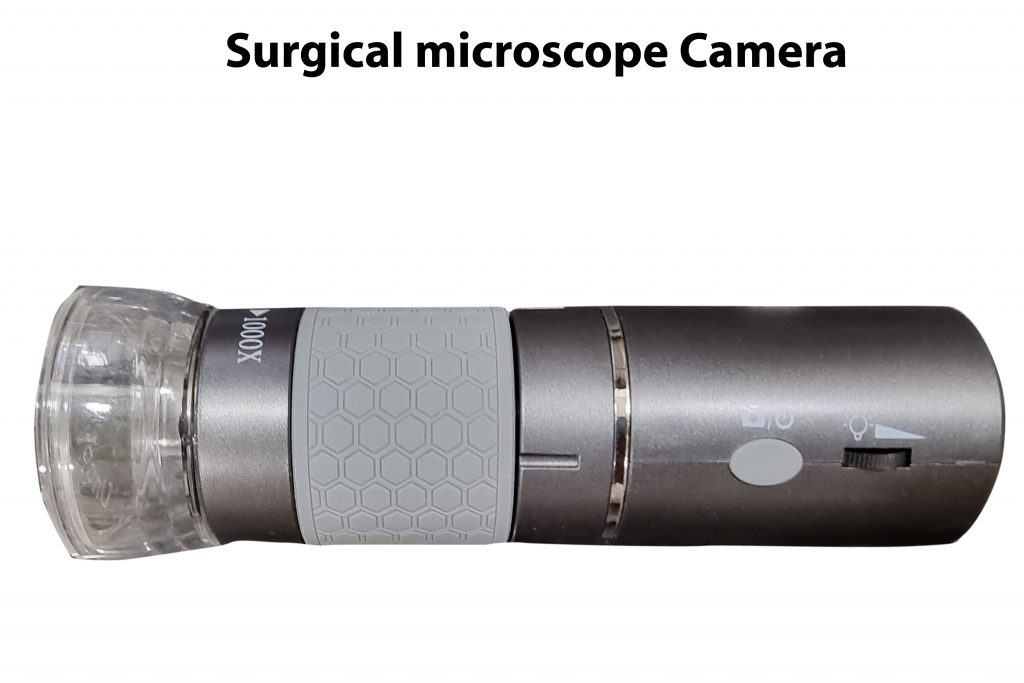
Product name: Surgical microscope Camera
Trade name: USB Microcam
Product Specifications: The Surgical microscope Camera has a video camera attached at the tip with lens assembly, lens holder, inner cylinder, focus ring, a video camera with 8 LEDs (light emitting diodes) for illumination and a stand for its holding and movement with a USB output. It has 200 X magnification with a side focus ring.

Detailed description: Complete specifications of the Surgical microscope Camera are: The device is tubular in shape and is made up of a lens assembly, lens holder, inner cylinder, focus ring, a video camera with LEDS for illumination and a stand for its movement. The stand has a base plate (figure:1B,13) with a vertical rod (figure:1B,9) with threads. (Figure:1B,12). A metal sleeve (figure:1B,10) moves up and down these threads (figure:1B,12) with 2 large screws. (Figure:1B,11) The sleeve has a bar with a ring (figure:1B,6) as a holder which holds the microscope. A small screw(figure:1B,5) fixes the microscope with the ring.

Product image
Figure:1A and B showing outer view of the Surgical microscope Camera
Figure:1A showing the front view of the tip of the Surgical Camera
1.Lens assembly 2. LEDS 3. Plastic cover
Figure:1B showing the outer side view of the Surgical microscope Camera with a stand
4.Focus ring 5. Screw for holding Surgical Camera 6. A ring holder 7. Button for taking still pictures 8. Output wire 9. Vertical rod of the stand 10. Sleeve that moves up and down 11. Wheels for the movement 12. Threads on vertical rod 13. Base of the stand

Figure:2 A, B, C showing the spare parts of the Surgical microscope Camera
Figure: 2A showing the top view of the focus ring
1.Focus ring 2. Groove for moving lens holder 3. Serrations for movement of focus ring
Figure:2B Top view of the inner cylinder of Surgical Camera
4. Groove for the movement of lens holder 5. Window for the focus ring 6. Inner cylinder of the Surgical CameraFigure:3C showing side view of the Surgical Camera7. Plastic cover 8. Main outer body of Surgical Camera 9. Focus ring 10. Button for taking still pictures 11. Wire with USB output

Figure:3 showing the inner view of the Surgical microscope Camera
1.Wire with USB output 2. Support for the metal rods 3. Video camera circuit plate 4. Metal rods 5. CMOS sensor of video camera 6. Outer body of Surgical Camera 7. Focus ring 8. Inner cylinder of Surgical Camera 9. Knob on lens holder which move in the groove of the focus ring 10. Knobs on lens holder which move in the grooves of inner cylinder 11. Lens holder 12. Groove in the focus ring 13. Cylinder containing lens assembly 14. LEDS 15. LED ring 16. Front plastic cover 17. Lens assembly
The device has an outer cylindrical shaped body. (Figure:1C,8) It has an inner cylinder (figure:2B,6 and figure:3,8) with groves(figure:2B,4) for the movement of the knobs of lens holder. Inside inner cylinder is a focus ring. (Figure:2A,1 and figure:2C,9 and figure:3,7) This focus ring (figure:2A,2) has groves from inside.
The front part of the microscope has a plastic cover (figure:1A,3 and figure:2C,7 and figure:3,16) to avoid outside light interfering in the microscope image. In the front part of the microscope, there is a ring (figure:3,15) with a central hole LEDS (figure:1A,2 and figure:3,14) for illumination.
The device has a cylindrical shaped structure containing lens assembly (figure:1A,1 and figure:3,17). It gives the it a reasonable magnification range. There is a lens holder (figure:3,10) at the bottom of this cylinder. The lens holder has 4 knobs (figure:3,9 and 10) placed diagonally opposite to each other, out of which the 2 move in the groove (figure:2B,4) of the inner cylinder over 2 metal rods and another 2 are moved by the groove (figure:2A,2 and figure:3,9) of the focus ring. As the focus ring moves, the lens holder and the lens assembly move forwards and backwards giving a focus range of 4 to 50 cm.
Behind the lens, these is a video camera (figure:3,3) with a CMOS chip. (Figure:3,5) The image of the lens falls on the CMOS chip and converted into a digital signal. The body of the microscope has a button (figure:1B,7) for taking still pictures. The camera has a wire with USB output with a switch for light control that controls the brightness of LEDs in the front. A USB wire connects the device to variety of devices such as desktop, laptop, android mobile phones and android based tabs. The microscope comes with software which needs to be installed on display devices to see and record the images and the video.
The technical advances of my invention are as follows. My Surgical microscope Camera has a tubular shape and is made up of a lens assembly, lens holder, inner cylinder, focus ring, a video camera with LEDs (light emitting diodes) for illumination and a stand for its holding and movement during surgery and a USB output. For operative recording, the distance of object from Surgical Camera should be 20 cm or more. There is image loss due to lenses. Due to Chip on the Tip technology, there is no image loss in my microscope. My microscope has a USB output as a male USB pin which connects to a USB cable that connects the device to the computer desktop, laptop, android device or a mobile phone and a free software given along the instrument installed on the display devices has a facility for display and recording of the video and images, a feature absent in prior art. My Surgical Camera can record a variety of operative procedures in Ophthalmology, Otolaryngology and in plastic surgery for microvascular surgery. Due to lack of above features, the prior art devices simply cannot be used as a replacement for my device. The prior art devices are imported and costly costing about Rs.2 Lakhs, whereas my device is cheap and indigenized and can be commercialized for only Rs. 30,000. My device is in tune with government of India’s Make in India policy.
Video quality: https://www.youtube.com/watch?v=5hK83hoeXGU
Intended application: The surgical microscope camera is used for recording a variety of operative procedures in general surgery, paediatric surgery, Ophthalmology, Otolaryngology and in plastic surgery for microvascular surgery. For this purpose, a separate stand is necessary to hold the Surgical Camera in various positions. The device can be used as a digital microscope for microvascular surgery in plastic surgery, for microvascular surgery of tubal recanalization and for Vas Difference recanalization, for ear and ophthalmic surgery etc.
Class A
Material: Plastic
Packing & storage Condition: The product is stored in a dust free cabinet at 25-40 % centigrade temperature. It is packed with multiple layers of bubble wraps and placed in a metal container of appropriate size and sent by courier
Cost: Rs.30,000


