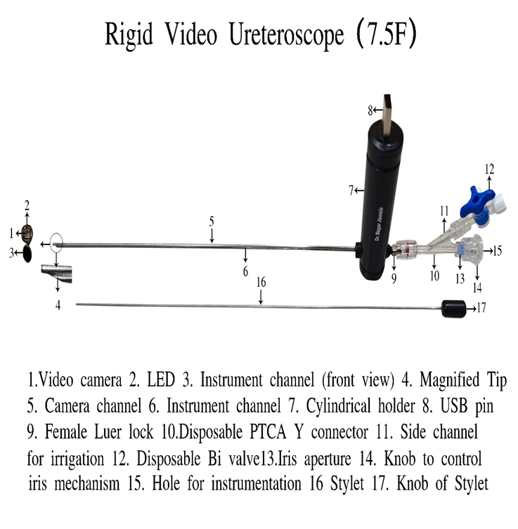
The Rigid Video Ureterorenoscope is characterised by two semirigid tubes fixed to each other, one below another with a holder at the middle end. The upper tube is the camera channel and the lower tube is the instrumentation cum irrigation channel. It has at the back end a female Luer lock. The device has a posterior disposable bridge which can be replaced easily. The bridge has a side channel for irrigation. A Bi valve fits to the side channel of the bridge to control water flow during the procedure. The bridge has a posterior end with an iris mechanism to control water flow. It has an outer diameter of 7.5 F and instrument channel of 4.5 F with a working length of 45 CM.
The upper tube is the camera channel and has a tubular color video camera with white light emitting diodes (LEDs) for illumination fixed at the tip thus does not require a separate light source as in traditional equipment. The lower tube of the device is a straight instrumentation channel that passes through the holder and ends in the posterior end as a female Luer lock. The same channel also acts as an irrigation channel for irrigation of fluid during endoscopy procedure. Fluid is passed through the side channel of the disposable bridge by attaching it to a disposable Bi valve. On other port of Bi valve, suction can be attached. The lever of the Bi valve can start and stop fluid passing to the tip of the scope into the body as well as can switch between suction and irrigation. Instruments are passed through instrumentation channel through a hole in the posterior knob, having an iris like washer. It avoids fluid leaks around the instrument; the fluid can pass around the instrument and the vision during endoscopy remains clear. Thus, the device has an integrated irrigation and suction system, allowing for simultaneous fluid management, thereby minimizing procedural interruptions and maintaining a clear field of vision.
It is extremely light weight with weight of 50 Gm compared to 500 Gm for the peers. The device is designed with a lightweight ergonomic structure ensuring ease of handling and reducing surgeon fatigue during prolonged procedures.
The device has a USB pin as output at the back end and a USB cable connects the device to a variety of devices such as desktop, laptop, android mobile phones and android based tabs and with a software installed on the display devices and the images and video can be seen as well as recorded on the display devises.
Cost Rs. 75,000
Enhancing Urological Precision – Beyond the Basics
You’ve already seen how the Rigid Video Ureteroscope is built from two semirigid tubes with an integrated camera channel and irrigation/instrumentation path – a design that keeps the view clear and the procedure smoother than older optics-only ureteroscopes.
What many clinicians notice when they start using this instrument is the way real-time video changes the feel of the surgery. The tubular colour video camera at the tip delivers a bright, consistent feed – it doesn’t rely on an external light source, because the LEDs are part of the scope itself. That simplicity isn’t just about convenience. It means the ureteroscope becomes an active part of decision-making in the OR.
In routine ureteroscopic work, the contrast to traditional fiber-optic rigs can be striking. Those older systems often required bulky light cables and separate camera heads, which can make set-up feel like a distraction. This USB-connectable design keeps the focus on the patient – plug into a laptop, tablet, or monitor, install the lightweight software and you’re visually tracking anatomy right away.
And while we’re talking of vision, it’s worth thinking about related tools you may already use. Like a Video Laryngoscope – originally engineered to let clinicians see around corners in the airway – the principle here is the same: don’t guess, see. That immediate visual feedback isn’t optional in delicate anatomy; it’s a baseline expectation. Ka-boom of clarity, no fumbling. That’s why video-based devices have reshaped so many fields of endoscopy.
It’s also useful to remember where a ureteroscope fits in the bigger picture of urological instrumentation. You might already be familiar with a Rigid Cystoscope on your equipment list – focused on the bladder and urethra, especially for diagnostic evaluations and interventions. The Rigid Video Ureteroscope takes those same high-definition realities and carries them deeper, up the ureter, toward the kidney.
Why Users Appreciate This Design
- Integrated video with illumination – no separate light source, no bulky cables, streamlined workflow.
- USB compatibility with common devices – real-time imaging and recording without specialized towers.
- Irrigation + suction control built into the bridge – so fluid management isn’t an afterthought.
- Lightweight ergonomics – around 50 g, which reduces fatigue during longer procedures.
- Clear field, fewer interruptions – the instrument channel’s design helps keep fluid from obscuring your view.
This isn’t just a ureteroscope that works. It’s a tool that reshapes the everyday experience of navigating upper urinary tract pathology. From stone evaluation to strictures and targeted biopsies, its video first approach keeps the focus where it should be: on what you see, rather than how you rig the camera.
Design and Construction
The Rigid Video Ureterorenoscope is characterised by two semi-rigid tubes fixed together, positioned one below the other and supported by a holder at the middle end. This structured alignment ensures stability and controlled handling during procedures. The upper tube functions as the camera channel, while the lower tube serves as the instrumentation cum irrigation channel. At the back end, the device is fitted with a female Luer lock connection for secure attachment.
A posterior disposable bridge is included and can be replaced easily. The bridge contains a side channel for irrigation where a Bi valve is attached to regulate water flow. The posterior end features an iris mechanism to control fluid movement precisely.
Technical Specifications:
- Outer diameter: 7.5 F
- Instrument channel: 4.5 F
- Working length: 45 cm
These specifications make this rigid ureteroscope suitable for standard ureteroscopic procedures.
Integrated Imaging System
The upper tube of the rigid ureteroscope contains a tubular colour video camera positioned at the distal tip. White LED lights are fixed alongside the camera, providing consistent illumination directly at the point of visualisation. This feature eliminates the need for a separate external light source as required in traditional systems.
Imaging Advantages:
- Built-in LED illumination
- No external light cable required
- Direct tip visualisation
- Stable real-time video output
The integrated imaging design of this rigid ureteroscope ensures immediate and reliable visualisation during procedures.
Instrumentation and Irrigation Channel
The lower tube functions as a straight instrumentation channel and extends to the posterior end where the female Luer lock is located. The same channel also acts as an irrigation pathway during the endoscopic procedure.
Fluid is delivered through the side channel of the disposable bridge using a Bi valve. The second port of the valve allows suction attachment when required. The valve lever enables controlled switching between irrigation and suction.
Instruments are introduced through a hole in the posterior knob fitted with an iris type washer. This design:
- Prevents fluid leakage around instruments
- Allows continuous fluid circulation
- Maintains a clear visual field
The integrated channel system allows this rigid ureteroscope to manage both instrumentation and fluid flow efficiently.
Integrated Fluid Management
The rigid ureteroscope is designed with a fully integrated irrigation and suction system, allowing simultaneous fluid management. Continuous irrigation helps maintain a clear operative field, while suction can be activated instantly when required.
Key Benefits:
- Smooth and controlled fluid flow
- Reduced procedural interruptions
- Improved visibility throughout the procedure
- Enhanced workflow efficiency
This coordinated system supports accurate and consistent surgical performance.
Ergonomic and Lightweight Design
Weighing approximately 50 grams, this rigid ureteroscope is significantly lighter than many conventional systems that may weigh around 500 grams. The lightweight construction improves manoeuvrability and reduces hand strain during prolonged procedures.
Ergonomic Features:
- Balanced grip design
- Reduced surgeon fatigue
- Better precision in delicate procedures
- Comfortable handling during extended use
The compact design enhances control while working in confined anatomical spaces.
USB Connectivity and Digital Recording
The rigid ureteroscope is equipped with a USB output pin at the back end. Using a standard USB cable, it can be connected to:
- Desktop computers
- Laptops
- Android mobile phones
- Android-based tablets
With compatible software installed, live images and videos can be viewed and recorded directly. This eliminates the need for bulky camera control units and simplifies operating room setup.
Clinical Applications
This rigid ureteroscope is suitable for various ureteroscopic procedures, including stone management, stricture evaluation, and diagnostic examination of the upper urinary tract.
Common Uses:
- Visualisation of ureteric stones
- Assessment of strictures
- Diagnostic ureteroscopy
- Evaluation of upper tract abnormalities
The combination of rigid stability, integrated video imaging, efficient fluid management, and lightweight handling makes this rigid ureteroscope a practical and reliable instrument for routine urological practice.
Cost
Cost: Rs. 75,000
This rigid ureteroscope combines structural strength, integrated imaging, ergonomic comfort, and digital connectivity to support precise and efficient urological procedures.