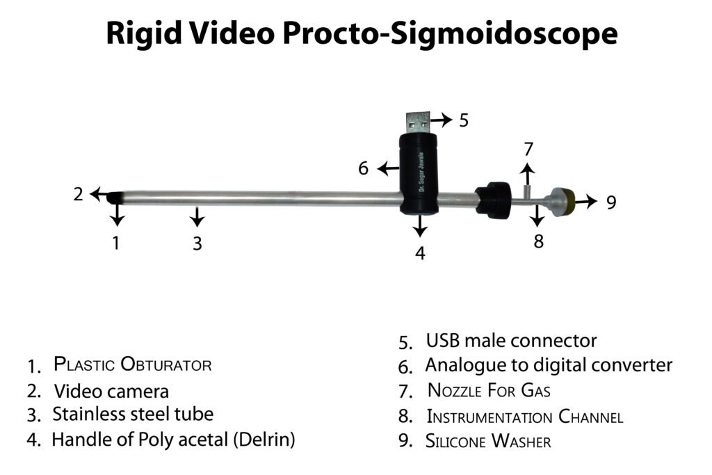
Product name: Rigid Video Procto-Sigmoidoscope 10,15 mm
Trade name: USB Proctocam 10, USB Proctocam 15
Product Specifications: The rigid video Procto-sigmoidoscope, a 5 in 1 device which is a merger of a telescope, light source, endo-camera, sheath and instrumentation channel. It is made up of a stainless-steel tube with a blunt obturator in front containing a video camera with 6 LEDs and electrical wires with an analogue to digital converter circuit placed in a plastic holder at the middle of the device with a USB pin as output with an instrumentation channel and a side port for gas or air insufflation. It has two sizes 10- and 15-mm diameter. It has zero-degree vision and 30 cm working length.
Detailed description: The rigid video Procto-sigmoidoscope is a 5 in 1 device which is a merger of a telescope, light source, endo-camera, sheath and instrumentation channel used for the purpose of proctosigmoidoscopy. The device is made up of a stainless-steel tube with a blunt obturator in front containing a video camera with LEDs and electrical wires with an analogue to digital converter circuit placed in a holder at the middle of the device with a male USB pin as output with an instrumentation channel and a side port for gas or air insufflation.
Proctosigmoidoscope or Rigid Sigmoidoscope
The Proctosigmoidoscope, also called as Proctoscope or rigid sigmoidoscope, is a thin but rigid tube fitted with a camera, light and lens. This is a five in one device. This tube is made of stainless steel. It’s a merger of a telescope, endo camera, light source, instrumentation channel and sheath. This device comes with a video camera and 6 LED lights. It is used by doctors to examine the rectum and the lower part of the large intestine (colon). It is gently inserted through the anus into the rectum. The name of this procedure is proctoscopy or rigid sigmoidoscopy. The rigid sigmoidoscope can be bent from the tip in four directions.
The Proctosigmoidoscope is also used to detect signs of hemorrhoids, cancer and polyps. This procedure helps in the diagnosis and prevention of various colon and rectal diseases.
Use of Proctosigmoidoscope, Rigid sigmoidoscope
Many diseases or conditions affect your rectum or colon. With the help of a proctosigmoidoscope, the doctor examines the rectum and the lowest part of the large intestine. The doctor can use the rigid sigmoidoscope , proctosigmoidoscope for the following reasons –
- To detect diseases in your rectum or colon
- To find the source of bleeding in the rectum
- To find the cause of diarrhea or constipation
- To stop the growth or development of existing polyps
- To check for colon cancer or to monitor previously treated rectal cancer
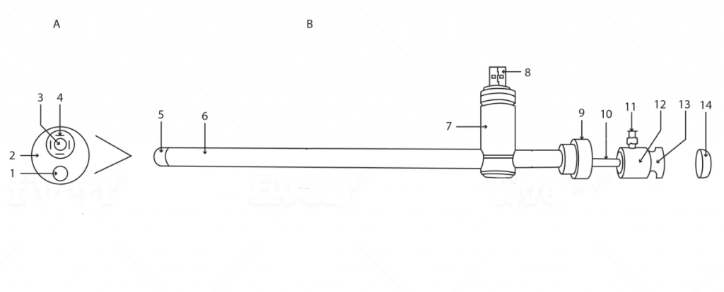
Figure: 1 A and B showing outer view of Rigid Video Procto-sigmoidoscope
Figure: 1A Front view of the tip of Rigid Video Procto-sigmoidoscope
1.Instrumentation channel 2. Obturator 3. Video camera 4. LEDs
Figure: 1B side view of Rigid Video Procto-sigmoidoscope
5. Side view of obturator 6. Stainless-steel tube 7. Cylindrical holder 8. Male USB pin 9. Round cap 10. Instrumentation channel 11. Nozzle with a Luer lock 12. Round hub 13. Groove for the silicone washer 14. Silicone washer
Complete specifications of Rigid Video Procto-Sigmoidoscope are as follows: It is made of a stainless-steel tube (figure:1B,6 and figure:2,5) with working length of 30 cm and outer diameter of 10 mm and 15 mm. The rigid instruments have a detachable obturator which is blunt which is put into sigmoidoscope before inserting into the anus to avoid injury to the patient. Since that is not possible in this instrument, this instrument is given a fixed and blunt round obturator as a tip. (Figure:1A 2,5 and figure:2,1) The video camera of is fixed at the upper end of this obturator. (Figure:1A,3 and figure:2,2). The video signal from the camera is carried by a bunch of wires (figure:2,4) through the metal tubes and connected to the analogue to digital circuit (figure:2,7) placed in the cylinder-shaped holder. (Figure:1B,7 and figure:2,6) The output of this circuit is given to the USB pin placed in the cylindrical holder. This holder is placed perpendicular to the stainless-steel tube. Behind the holder, there is a round cap (figure:1B,9 and figure: 2,9) This cap gives support to the main stainless-steel tube. The instrumentation channel passes through this cap through a small hole. A USB wire connects the device to variety of devices such as desktop, laptop, android mobile phones and android based tabs. The device comes with software which needs to be installed on the display devices to see and records the images and the video. The video camera has LEDs (figure:1A,4) which are powered by the power from USB pin. The LEDs illuminate the field during the procedure. The device has an instrumentation channel fixed in front at the lower end of the Delrin obturator. (Figure:1A,1 and B,10 and figure:2, 3,12) It has an inner diameter in the range of 0.1 mm to 15 mm that accommodates a variety of instruments. The back of the instrument channel is in a round hub. (Figure:1B,12 and figure:2,10). This round hub has a groove (figure:1B,13) Over this groove attaches a silicone washer (figure:1B, 13) so that the air or gas should not leak during the procedure. At the side of the Delrin hub, there is a side channel with a nozzle with a Luer lock (figure:1B,11 and figure:2,11) which is welded to the main instrumentation channel. Thus, gas channel and instrumentation channel are common. The gas channel allows for gas insufflation during the procedure. To this channel, a balloon of BP apparatus may be attached for air insufflation. Alternatively, gases such as O2 or Co2 can also be connected with a tubing.
Parts of the Proctosigmoidoscope
The Proctosigmoidoscope is about 30 cm long and has zero degree vision. It has a camera and a light on the tip. The main part of this instrument is cylindrical and is made of stainless steel. It is hollow so that the rectum can be cleaned properly. It also has a side port for gas or air insufflation. The Rigid Sigmoidoscope can be used more than once. Its USB wire can be connected to any laptop, desktop, android mobile devices and tabs. The instrument comes with a software that you will need to install.
The handle is ergonomically designed to make it easy to hold and use the proctosigmoidoscope. The tip is smooth and rounded so that it can be inserted comfortably into the rectum without causing any bruising. There is also a light at the end of the instrument that illuminates the walls of the rectum and colon so that the doctor can see clearly.
How the proctosigmoidoscope is used
Your doctor will ask you to undress from the waist down and lie on your left side with your knees bent on a table. The doctor will first perform a preliminary digital rectal examination with gloved fingers. This test checks for blockage or tenderness of the rectum. After this examination, he will use the lubricated rigid sigmoidoscope and gently insert it into your rectum. You may feel uncomfortable during this process and feel like cramping but this is quite normal. This test does not cause any kind of pain but if you feel like you are afraid of pain then you can ask the doctor for anesthesia. Proctosigmoidoscopy will only take about 5 to 15 minutes.
What happens after proctosigmoidoscopy
After proctosigmoidoscopy, most people get busy with their normal work. If your doctor has used air in your rectum, then you may feel bloating or gas. You are likely to have light bleeding 1 or 2 times during the toilet, often this is due to Biopsy.
Who can use the Proctosigmoidoscope
Only gastroenterologists and colorectal surgeons can use the Rigid Sigmoidoscope. Proctosigmoidoscope is used to examine the anus and rectum. The doctor can remove some small tissue samples during the Proctosigmoidoscopy.
Proctosigmoidoscope vs other scopes
Rigid Sigmoidoscope or Proctoscope: It only examines the rectum and anus
Anoscope: It is smaller than the proctoscope and is used only to examine the anus.
Flexible sigmoidoscope: It is a long flexible tube that goes into the rectum and colon.
Colonoscope: It is the longest flexible scope that examines the large intestine.
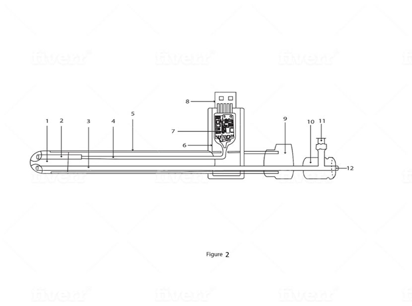
Figure:2 showing the inner view of Rigid Video Procto-sigmoidoscope
1.Obturator 2. Video camera with LEDs 3. Instrumentation channel 4. Electrical wires 5. Outer stainless-steel tube 6. Holder 7. Analogue to digital converter circuit 8. Male USB pin 9. Round cap 10. Round hub 11. Nozzle with a Luer lock 12. Back end of instrumentation channel
The output of this circuit is given to the USB pin. A USB wire connects the device to variety of devices such as desktop, laptop, android mobile phones and android based tabs. The device comes with software which needs to be installed on the device to see and records the images and the video.
The device is reusable and can be sterilized by putting in Formalin chamber for 30 minutes or by Ethylene Oxide gas.
The advantages of the device over the conventional are as follows: It is a compact and lightweight device which is 5 in 1 in the nature and is a unique merger of a Telescope,
light source and endo-camera, sheath and instrumentation channel. It is commercialized at a reasonable cost of Rs.40,000 compared to Rs. 6 Lakhs of the conventional equipment. The instrument can perform a lot of endoscopic surgeries such as endoscopic biopsy for Hirschsprung’s disease and operative procedures such as polypectomy, coagulation of rectal ulcers, banding of piles etc. which are difficult for the conventional instrument. The picture quality is excellent due to the “Chip on Tip” technology and there is no image loss as in conventional instrument.
Intended application: It can do variety of diagnostic procedures such proctoscopy, sigmoidoscopy and therapeutic procedures such as endoscopic biopsy for Hirschsprung’s disease and operative procedures such as polypectomy, coagulation of rectal ulcers, banding of piles etc.
Method of using: The device is sterilized by Ethylene Oxide gas sterilization. The patient in lithotomy position with parts prepared and draped. The device is connected via a USB cable to the display devises and images and videos start streaming on them. The device is gently inserted into rectum and the side gas nozzle connected to C02 insufflator with 10 cm of water pressure and 10 liters per minute flow. The illumination of LEDs adjusted to get a clear picture. The recta lesion identified clearly. A biopsy forceps passed through the instrumentation channel to get the biopsy of the lesion. Many operating forceps can be passed through instrumentation channel for various surgeries. Images and video recorded on a software installed on display devises. The device gently taken out and cleaned and sent back for sterilization.
Class B
Material: 316 grade stainless steel
Patent no: 202021022106
Packing & storage Condition: The product is stored in a dust free cabinet at 25-40 % centigrade temperature. It is packed with multiple layers of bubble wraps and placed in a metal container of appropriate size and sent by courier
Video quality: https://www.youtube.com/watch?v=baRbg10aLwE&t=2s
Cost: Rs.40,000
Visual Feedback That Changes How You Examine the Lower Bowel
A procto sigmoidoscope may look simple in form, but the experience of using it depends heavily on what you can actually see once the examination begins. The Rigid Video Practo Sigmoidoscope brings the camera and illumination directly into the scope, creating a steady visual field through the rectum and sigmoid colon. Instead of relying on indirect views or frequent repositioning, the image remains consistent as the scope advances. This helps clinicians stay focused on tissue assessment, mucosal patterns, and small abnormalities that can otherwise be overlooked during routine examinations.
- Integrated video system keeps illumination aligned with the viewing field throughout the examination
- Real time visual output supports direct observation and straightforward documentation on common display devices
- Rigid construction allows controlled navigation and stable imaging without unnecessary flex or drift
- Familiar handling enables an easy transition from conventional rigid scopes with minimal adjustment
- Clean imaging supports clearer identification of lesions, inflammation, or areas requiring biopsy
When viewed alongside instruments such as a Rigid Esophagoscope, the value of direct video imaging becomes even clearer. In both upper and lower endoscopy, clarity reduces hesitation. Subtle mucosal changes are easier to interpret when lighting and focus remain stable. Over repeated use, this reliability supports more confident diagnostic decisions and smoother patient interactions, particularly in outpatient and screening environments where efficiency and accuracy both matter.
Clinical Clarity That Fits Into Real Practice
On paper, the Rigid Video Procto-Sigmoidoscope reads like a neat five-in-one solution. In real use, it feels more like a tool that simply stays out of the way. Once inserted, the image settles quickly and does not keep demanding attention. The camera and LEDs at the tip give a clear, evenly lit view of the rectum and sigmoid area, without the constant micro-adjustments that slow things down. For routine examinations, that steadiness matters. It lets the clinician move forward naturally, focus on the mucosa, and notice changes without breaking the flow of the exam.
What also becomes obvious is how predictable the handling feels. The rigid stainless-steel body guides movement, the obturator keeps entry smooth, and the visual feedback matches what the hands are doing. There is no delay, no sudden loss of brightness, no guesswork about where the scope is pointing. Over time, this consistency builds confidence, especially during busy outpatient sessions where efficiency and accuracy need to coexist.
A Shared Approach With Other Rigid Endoscopes
If you have worked with instruments like a Rigid Bronchoscope, the logic behind this design will feel familiar. Rigid scopes rely on control and direct vision, not flexibility or complex mechanics. The same thinking applies here. The moment the image appears on screen, it becomes easier to orient yourself and proceed calmly. Small lesions, early inflammatory changes, or bleeding points stand out more clearly when the view does not shift unexpectedly. The USB video output also adds to that ease. Images and videos can be viewed on common devices and recorded without additional equipment.
Insufflation through the side port and instrument passage through the working channel happen smoothly, without interrupting the examination. After a few cases, the device stops feeling new and starts feeling reliable, which is usually when equipment proves its worth. In everyday practice, this scope does not try to impress. It supports examination quietly, keeps the visual field stable, and allows clinicians to work at their own pace. That balance is what makes it useful beyond its specifications.