Flexible Video Ureteroscope is a sophisticated medical device that revolutionized the diagnosis and treatment of upper urinary tract diseases. Specifically created for direct viewing and treatment of sites like the ureters and kidneys, this highly flexible scope is a key tool in minimally invasive disease management of the urinary tract, such as kidney stones, stricture, tumors, and upper tract urothelial carcinoma.
Due to its low-profile design, digital imaging potential, and enhanced articulation, the Flexible Video Ureteroscope is a standard tool in the urology clinics of the world today.
The Flexible Video Ureteroscope is designed with great care for its ability to move through the delicate, intricate anatomy of the renal pelvis and the ureter with less tissue trauma.
Instruction video
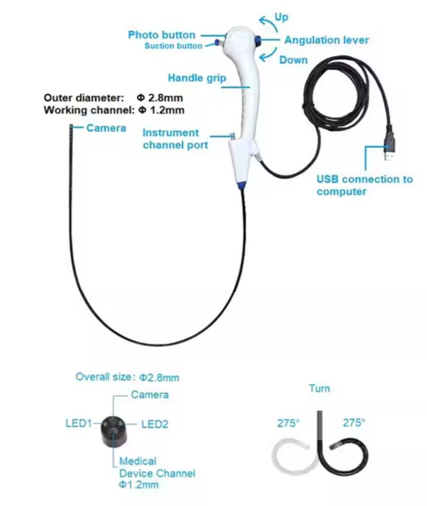
Structural Key Elements
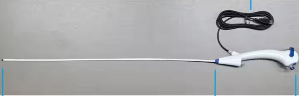
Insertion Tube: The instrument contains a thin, extremely pliable shaft—usually 7.5 to 9 French in size—making it easy to and atraumatically pass through the urinary tract. It makes preoperative dilation of the ureter unnecessary in the majority of cases.
Tip Articulation: The distal tip of the scope can be deflected two or four ways to a distance of as much as 270 degrees. This gives access to all areas of the kidney, even the frequently difficult-to-reach lower pole calyces.
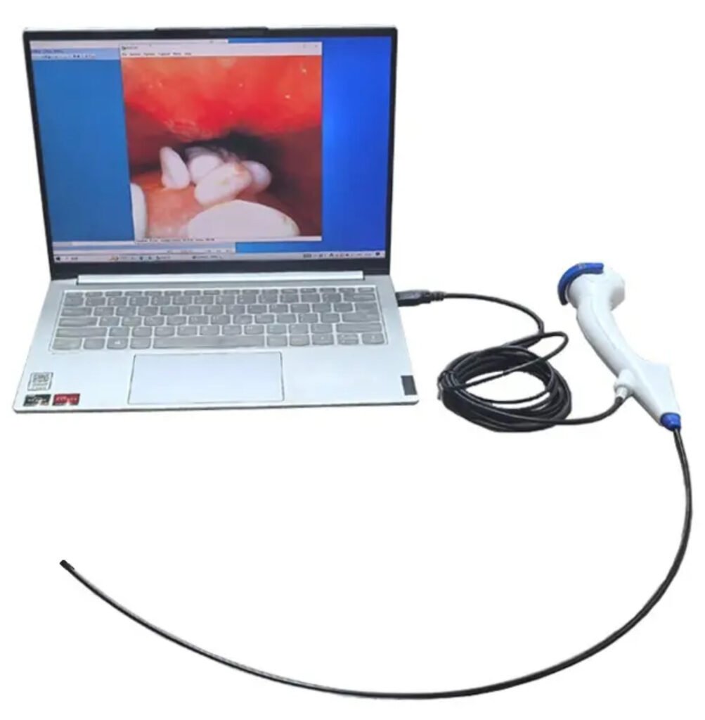
Imaging System: In contrast to the conventional fiberoptic scopes, the Flexible Video Ureteroscope contains a digital sensor, i.e., a CMOS or CCD chip, on its tip. This system can perform real-time, high-resolution imaging, significantly enhancing visualization during the procedure.
Working Channel: The integral working channel (typically ≤ 3.6 Fr) has space for a variety of endoscopic instruments like laser fibers, retrieval baskets, and biopsy forceps that can be used for both diagnosis and therapy.
Irrigation Channels: The series features special irrigation channels that provide an open operating field by flushing the field with sterile fluid intermittently or continuously.
Key Functional Features
Flexible Video Ureteroscope has numerous benefits that render it more effective in application in the clinic:
Digital Visualization: The high-definition visualization provides clear images of the urinary tract, allowing urologists to spot small stones, subtle tumors, or mucosal lesions missed by lower-resolution technology.
Improved Navigation:The flexible tip and the ergonomic control handle enable close control over the scope, making it simple for the operator to explore and examine every crevice of the renal collecting system.
Minimally Invasive Access: The Flexible Video Ureteroscope procedure is most often done without any incision at all, using general anesthesia. Patients have less postoperative pain, quicker recovery times, and fewer hospital days than with open or percutaneous methods.
Reusable and Disposable Models: Single use and reusable options are both available, allowing institutions to select on the basis of budget, infection control, and volume of cases.
Clinical Uses of the Flexible Video Ureteroscope
The Flexible Video Ureteroscope is a useful tool in diagnosis and therapy of most upper urinary tract pathologies.
The most frequent use is in stone management. Urologists utilize the laser fibers, e.g., Holmium: YAG or Thulium fiber lasers, inserted via the working channel to shatter and extract stones.
Direct visualization and biopsy of the renal pelvis or ureters’ suspected malignant lesions are possible through the procedure, facilitating diagnosis of upper tract urothelial carcinoma.
Management of Ureteral Stricture: The instrument allows safe dilatation or incision of strictures under direct vision, enhancing safety and success.
Foreign Body Removal: Foreign bodies or stents migrated into the urinary system can be removed safely with baskets or graspings passed through the working channel.
Postoperative Surveillance: Previously treated patients with stone or tumor can be surveyed with follow-up studies utilizing the Flexible Video Ureteroscope to survey for recurrence or complications.
Advantages Over Fiberoptic Scopes
The Flexible Video Ureteroscope provides several advantages over traditional fiberoptic ureteroscopes:
- Better Image Quality: Digital sensors provide brighter, sharper images, enhancing diagnostic quality and surgical accuracy.
- Greater Stable Imaging Long-Term: In contrast to fiberoptic systems, digital sensors are immune to image deterioration due to breakage or wear of the fibers.
- Better Ergonomics: Less weight and better-balanced scopes minimize surgeon fatigue and enable smoother, more controlled movement for longer procedures.
- Limitations and Considerations: While highly beneficial, the Flexible Video Ureteroscope suffers some drawbacks:
- Higher Upfront Costs: Digital and disposable scopes cost more upfront than fiberoptic, though cost savings should eventually increase with lower maintenance and greater performance.
- Reuse Models Maintenance: Reusable models are subject to sterile cleaning, disinfection, and handling procedures to maintain performance and avoid infection risk. The tip of high-tech and integrated camera systems should be handled with care to prevent damage.
Future Directions of Ureteroscopy
The future of Flexible Video Ureteroscope is bright with continued development being pursued towards making it more useful:
- Miniaturization; Wider scopes will enable treatment in smaller anatomy, i.e., pediatric cases or highly strictured ureters.
- Artificial Intelligence Integration: Diagnoses with AI may even be able to detect lesions or assist easier passage through procedures.
- 3D and Advanced Imaging: Greater resolution of images, 3D imaging, and augmented reality overlays may further enhance accuracy.
Robotic Assistance: The robotic control systems should enhance precision in movements and minimize human error, particularly in complicated situations.


Disclaimer: Please sterilise by putting in Formalin chamber for 30 minutes by ETO gas. Do not autoclave, do not put in Cidex. Clean thoroughly after use, flush instrument channel with 10 ML 1% betadine solution and rinse afterwards with 10 ML NS. Place protective sleeve provided over the Distel end. Put in same metal box after use in bubble wrap or in a plastic box.