The Rigid Video Esophagoscope is a new medical miracle used for exploring the esophagus more precisely, more efficiently, and at reduced expenses. The equipment has been in rising demand in ENT and gastroenterology because of its portability, affordability, and simplicity. As opposed to the conventional equipment utilized in esophagoscopy, the equipment provides high-quality video imaging in portable, combined form.

Product name: Rigid Video Esophagoscope, 8 mm
Trade name: USB Esocam 8
Product Specifications: The rigid video esophagoscope, a 5 in 1 device which is a merger of a telescope, light source, endo-camera, sheath and instrumentation channel. It is made up of a stainless-steel tube with a blunt obturator in front containing a video camera with 6 LEDs and electrical wires with an analogue to digital converter circuit placed in a plastic holder at the middle of the device with a USB pin as output with an instrumentation channel and a side port for gas or air insufflation. It has 8 mm diameter and has zero-degree vision and 30 cm working length.
What is a Rigid Video Esophagoscope?
A Rigid Esophagoscope is a straight, rigid tubular instrument constructed of hardened stainless steel for direct visualization and uncomplicated procedures in the esophagus. It is a 5-in-1 multidiagnostic endoscope that integrates a telescope, light source, endo-camera, sheath, and instrumentation channel into one.
About 30 to 40 cm in length, the design of the Rigid Esophagoscope is with strength and accuracy in mind. With its stainless steel tube, the esophagoscope is equipped with an obturator that has a high-resolution video camera and six LED lights for the best internal light. What’s special about this device is its advanced bringing together of technology, minimizing the need for multiple separate instruments in a procedure.

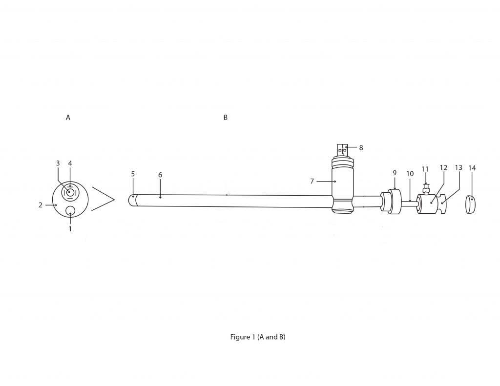
Figure: 1 A and B showing outer view of Rigid Video Esophagoscope
Figure: 1A Front view of the tip of pediatric size Rigid Video Esophagoscope
1.Instrumentation channel 2. Delrin obturator 3. Video camera 4. LEDs
Figure: 1B side view of pediatric size Rigid Video Esophagoscope
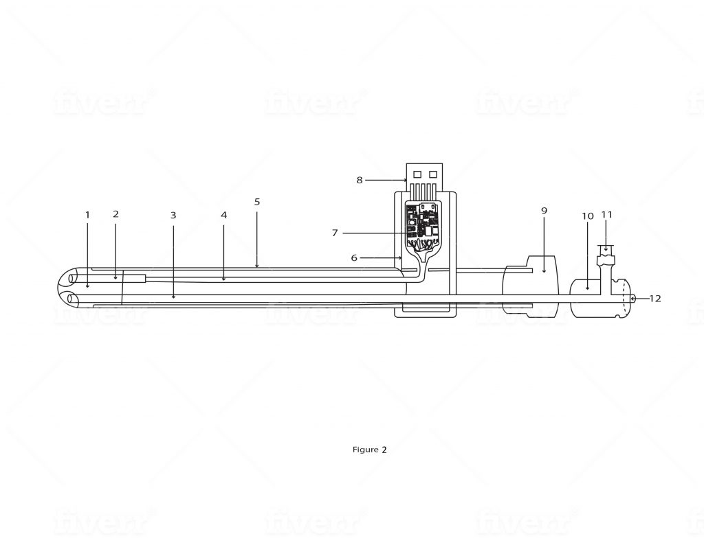
Detailed description: The rigid video Esophagoscope is a 5 in 1 device which is a merger of a telescope, light source, endo-camera, sheath and instrumentation channel used for the purpose of esophagoscopy. It is made of a stainless-steel tube (figure:1B,6 and figure:2,5) of outer diameter of 8 mm and working length of 40 cm. The rigid instruments have a detachable obturator which is blunt which is put into sigmoidoscope before inserting into the esophagus to avoid injury to the patient. Since that is not possible in this instrument, this instrument is given a fixed and blunt round plastic tip (figure:1A 2,5 and figure:2,1) made up of heat-resistant plastic called Delrin (polyacetal). A video camera of 4 mm diameter and 22 mm length is fixed at the upper end of this Delrin obturator. (figure:1A,3 and figure:2,2). The video signal from the camera is carried by a bunch of 10 wires (figure:2,4) through the metal tubes and connected to the analogue to digital circuit (figure:2,7) placed in the cylinder made up of heat resistant plastic called Delrin (Polyacetal). (figure:1B,7 and figure:2,6) The output of this circuit is given to the male USB pin placed in the Delrin cylinder. This Delrin cylinder is placed perpendicular to the stainless-steel tube. Behind the Delrin cylinder, there is a round cap (figure:1B,9 and figure:2,9) made up of Delrin and has a diameter of 25 mm. This cap gives support to the main 8 mm stainless-steel tube. The instrumentation channel passes through this cap through a small hole. A USB wire connects the device to variety of devices such as desktop, laptop, android mobile phones and android based tabs. The device comes with software which needs to be installed on the display devices to see and records the images and the video. The video camera has 4 LEDs (figure:1A,4) which are powered by the power from USB pin. The LEDs illuminate the field during the procedure.
Structure and Design Features
The Rigid Esophagoscope’s most striking feature is its look. A series of wires is carried through a plastic box situated in the center inside the stainless steel tube. They are connected to an instrumentation channel and a USB outlet for the supply of gas or air during the procedure.
In rigid instruments used traditionally, the obturator can be taken out for safety. In the Rigid Esophagoscope, however, the obturator is permanent, and a plastic tip rounded off and Delrin—a heat-resistant plastic used in medicine—is placed on the distal end for safe patient application. The plastic tip prevents tissue trauma on insertion and manipulation.
The camera is supported on a string of ten wires which pass through the steel tube and end in a Delrin cylinder, the USB output also being contained within it. The 8 mm diameter tube is supported by the Delrin cap at the end of the cylinder. The USB interface is plug-and-play compatible with monitors, laptops, Android phones, or tablets, and live viewing and recording of the procedure are facilitated with special software.

5. Side view of Delrin obturator 6. Stainless-steel tube of 10 mm diameter 7. Cylindrical holder of Delrin9 polyacetal) 8. Male USB pin 9. Round cap of Delrin 10. Instrumentation channel 11. Female Luer loch type nozzle 12. Round hub of Delrin 13. Groove for the silicone washer 14. Silicone washer
Figure: 2 A and B showing outer view of Rigid Video Esophagoscope
Figure: 2 A Front view of the tip of pediatric size Rigid Video Esophagoscope
1.Instrumentation channel 2. Delrin obturator 3. Video camera 4. LEDs
Figure: 2 B side view of pediatric size Rigid Video Esophagoscope
5. Side view of Delrin obturator 6. Stainless-steel tube of 10 mm diameter 7. Cylindrical holder of Delrin9 polyacetal) 8. Male USB pin 9. Round cap of Delrin 10. Instrumentation channel 11. Female Luer loch type nozzle 12. Round hub made of Delrin 13. Groove for the silicone washer 14. Silicone washer
The device has an instrumentation channel fixed in front at the lower end of the Delrin obturator. (figure:1A,1 and B,10 and figure:2, 3,12) It has a 2.5 mm inner diameter that accommodates a variety of 2 mm instruments. The back of the instrument channel is in a round plastic hub made up of Delrin plastic. (figure:1B,12 and figure:2,10). This round hub has a groove (figure:1B,13) Over this groove attaches a silicone washer (figure:1B,13) so that the air or gas should not leak during the procedure. At the side of the Delrin hub, there is a side channel with a female Luer lock (figure:1B,11 and figure:2,11) which is welded to the main instrumentation channel. Thus, gas channel and instrumentation channel are common. The gas channel allows for gas insufflation during the procedure. To this channel, a balloon of BP apparatus may be attached for air insufflation. Alternatively, gases such as O2 or Co2 can also be connected with a tubing.
The output of this circuit is given to the male USB pin. A USB wire connects the device to variety of devices such as desktop, laptop, android mobile phones and android based tabs. The device comes with software which needs to be installed on the device to see and records the images and the video. The device is reusable and can be sterilized by putting in Formalin chamber for 30 minutes or by Ethylene Oxide gas.
The advantages of the device over the conventional are as follows: It is a compact and lightweight device which weighs only 120 gm. It is 5 in 1 in the nature and is a unique merger of a Telescope, light source and endo-camera, sheath and instrumentation channel. It is commercialized at a reasonable cost of Rs.40,000 compared to Rs. 6 Lakhs of the conventional equipment. The picture quality is excellent due to the “Chip on Tip” technology and there is no image loss as in conventional instrument.
- Light and Slim Design: The Rigid Esophagoscope weighs only 120 grams, making it very light and convenient to maneuver. The fact that several functionalities are all integrated into one tool not only saves space but also makes the whole process flow throughout procedures simpler.
- Affordable Price: Another major benefit of this machine is its affordability. While standard endoscopic instruments vary between INR 6 lakh and above, the Rigid Video Esophagoscope is available for merely INR 40,000, which is affordable for small clinics, schools, and developing areas.
Chip-on-Tip Technology: With the newest chip-on-tip technology, the device provides better images and videos. Unlike earlier models, in which image quality would decline with use, the Rigid Esophagoscope provides consistent picture quality, allowing for accurate diagnosis and documentation.
Intended application: The instrument can perform a variety of diagnostic procedures such as diagnostic esophagoscopy for strictures, foreign bodies, oesophageal varices and therapeutic procedures such as biopsies, oesophageal variceal therapy and dilatation of oesophageal strictures etc.
Class B
Method of using the device: The device is sterilized by Ethylene Oxide gas sterilization. The patient in lithotomy position with parts prepared and draped. The device is connected via a USB cable to the display devises and images and videos start streaming on them. The device is gently inserted into rectum and the side gas nozzle connected to C02 insufflator with 10 cm of water pressure and 10 Liters per minute flow. The illumination of LEDs adjusted to get a clear picture. The recta lesion identified clearly. A biopsy forceps passed through the instrumentation channel to get the biopsy of the lesion. Many operating forceps can be passed through instrumentation channel for various surgeries. Images and video recorded on a software installed on display devises. The device gently taken out and cleaned and sent back for sterilization.
Uses and Clinical Applications
The Rigid Esophagoscope is employed in several diagnostic and intervention procedures of the esophagus. Some of its typical uses are:
- Detection of tumors and stricture of the esophagus
- To take biopsies
- For foreign body removal
- Esophageal dilatation procedures
- Endoscopic treatment of focal conditions
- Its rigid structure allows more internal space than flexible scopes, allowing greater instrument access and control with procedures.
How the Procedure is Performed
The Rigid Esophagoscope is inserted after the patient has received general anesthesia to numb the esophagus and avoid causing pain. The physician inserts the scope into the mouth and slowly into the esophagus. A video camera sends images from the scope in real time to a monitor, which assists the physician in viewing narrowing, inflammation, or tumors.
Depending on the level of complexity of the condition, the process lasts from 30 minutes to 3 hours. Minor side effects such as a dry throat may follow the process but clear up readily.
Maintenance and Sterilization
The Rigid Esophagoscope requires regular cleaning and maintenance for it to be long-lasting and hygienic. After every use:
- The components are disassembled with caution.
- Equipment is cleaned using an enzymatic cleanser.
- It is thoroughly washed with clean water.
- Air drying is performed in order to eliminate moisture.
- The main elements are sterilized prior to storage or reuse.
- These steps contribute to instrument performance and avoid cross-contamination.
Why purchase the Rigid Video Esophagoscope
Vigyan Yog’s Rigid Esophagoscope is a cost-efficient, technologically advanced, and versatile gadget that has made it possible to treat and diagnose the esophagus. Its easy handling, light weight, and excellence in image quality have turned it into a highly sought-after gadget for contemporary medical facilities. For identifying tumors, biopsy, or the removal of obstructions, this gadget offers quality and guaranteed performance. With advancements in technology, innovations such as the Rigid Esophagoscope are leading to improved and safer patient care in endoscopy.
Patent No: 202021022102
Material: 316 grade stainless steel
Packing & storage Condition: The product is stored in a dust free cabinet at 25-40 % centigrade temperature. It is packed with multiple layers of bubble wraps and placed in a metal container of appropriate size and sent by courier
Video quality: https://www.youtube.com/watch?v=FQqGjHP6wuY
Cost: Rs.40.000


