When accuracy in airway access, visualization, and intervention is paramount, there’s one instrument that continues to justify its place at the top: the rigid bronchoscope. With ubiquitous application across pulmonary medicine, thoracic surgery, and emergency airway management, this instrument of excellence is a highly valued asset in the arsenal of any hospital or surgical center.

Rigid Ventilating Video Bronchoscope
Product name: Rigid Video Bronchoscope with forcepsTrade name: USB Bronchocam F
Product Specifications: Rigid ventilating video bronchoscope with forceps, is a device with “Chip on the tip” technology, a 5 in one device, a merger of telescope, light source, ventilating sheath, endo-camera, and forceps, comprising of a ventilating sheath of bronchoscope with side holes for ventilation in front with a peanut holding forceps and handle with a video camera and 4 LEDs (Light emitting diodes) fixed at the tip of the sheath, a ventilation port at the back end and a holder with a USB pin as output. It has zero-degree vision and 40 cm working length.
Detailed description: Complete specifications of rigid ventilating video bronchoscope with forceps: It is a device with “Chip on the tip” technology, a 5 in one device, a merger of telescope, light source, ventilating sheath, endo-camera, and forceps, comprising of a ventilating sheath of bronchoscope with side holes for ventilation in front with a peanut holding forceps and handle with a video camera and LEDs (Light emitting diodes) fixed at the tip of the sheath, a ventilation port at the back end and a holder with a USB pin as output used for a medical procedure called bronchoscopy.
What is a Rigid Bronchoscope?
Rigid bronchoscope refers to a stainless steel tube employed by thoracic surgeons and pulmonologists in order to visualize and manage airway diseases. Rigid bronchoscopes are largely used for interventional as well as diagnostic procedures, as opposed to flexible bronchoscopes, which are mainly diagnostic.
It is inserted into the patient’s mouth and offers quick access to the trachea and major bronchi. Light and telescope are inserted into the tube, giving an excellent, lighted image of the airway on a monitor or directly through eye observation of the eyepiece. It even allows the exchange of devices like forceps, suction catheters, and stents—rendering it an all-in-one solution for instant airway procedure.
Why Should a Rigid Bronchoscope Be Used?
While flexible bronchoscopes certainly have their role in modern medicine, there are certain clinical situations where the rigid bronchoscope proves to be especially useful. Some of them are:
- Better airway control in emergency situations
- Better blood, secretion, or debris suctioning
- Debulking of bulky foreign bodies
- Increased working channel diameter for instrumentation
- Facilitating laser treatment, stenting, and tumor debulking
If your procedure includes therapeutic bronchoscopies or airway surgery, rigid bronchoscope is a must-have.
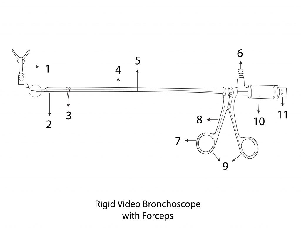
Figure 1 showing the outer view of the rigid ventilating video bronchoscope with forceps
1.Peanut holding forceps 2. Video Camera 3. Holes for ventilation 4. Ventilating channel 5. Tube of forceps 6. Port for ventilation 7. Rings of handle of the forceps 8. Arms of handle 9. Handle of forceps 10. Holder 11. USB pin
The main ventilating channel of the device is a hollow tube (figure:1-4, figure 2-7) which has holes (figure:1-3, figure 2-6) on each side for ventilation in front and a ventilating side port (figure:1-6, figure 2-11) at the back end. A video camera (figure:1-2 and figure:2-2) with LEDs (Figure:2-3) is fitted at the tip of the main ventilating channel. Electrical wires (figure: 2-5) bundled into a single core pass through the ventilating channel and is connected to another end of the instrument to an analogue to digital converter circuit (figure:2-14) placed in a hollow holder. (Figure:1-10 and figure:2-15) The device has a peanut holding foreign body removal forceps (figure:1-1 and figure:2-1) welded below the main ventilating channel. The forceps has a handle at the back end with arms (figure:1-8 and figure:2-12) and rings (figure:1-9 and figure:2-13) for placing fingers. The body of forceps (figure:2-8) has a wire (figure:2-8) that passes through the body and attached to the hinges at the jaws of the forceps (figure:1-1) in front. It attaches to the backend to the head of the arms (figure:2-10) of the handle of the forceps. Thus, the jaws of the instrument can be closed by approximating the arms of the handle towards each other and opened by moving the arms in opposite direction. The back end of the device has a hollow cylindrical holder (Figure:1-10 and figure:2-15) which accommodates analogue to digital circuit of the front camera and has a USB pin as output (Figure:1-11 and figure:2-16). A USB cable connects the instrument to the computer desktop, laptop, android device or a mobile phone and a free software given along the instrument has a facility for display and recording.
The instrument is reusable, light weight (150 gm) and is sterilized by putting in Formalin chamber for 30 minutes or by Ethylene Oxide gas. There are no fiber optics and rod lenses, hence no image loss excellent image quality and the instrument can record still images and video from its inbuilt camera without the need of external camera. The device is used for a variety of diagnostic bronchoscopic procedures such as diagnosis of tracheoesophageal H type fistula, medical lung diseases and can also be used for therapeutic bronchoscopy for removal of bronchial foreign body, biopsy of suspicious lesions, brush biopsy and for bronchial lavage, etc.
Top Advantages of a Rigid Bronchoscope
Optimal Optical Clarity: Our rigid bronchoscopes are fitted with state-of-the-art telescopic technology and high definition optics, offering an excellent, lighted view of the tracheobronchial tree. With either camera system or eyepiece in use, there is clear and consistent visualization.
Large Instrument Channels: The spacious inner lumen of the rigid bronchoscope is convenient to use with an assortment of instruments such as grasping forceps, electrocautery probes, suction devices, and laser fibers—i.e., extremely convenient in therapeutic procedures.
Stainless Steel Construction Which Would Endure: Made of finest quality medical stainless steel, the rigid bronchoscope can be autoclaved, is corrosion-proof, and can be reused multiple times. This renders it economical for clinics and hospitals.
Simple to Clean and Maintain: The simple, modular construction allows quick disassembly and cleaning between cases. Infection control is important, and our bronchoscopes are constructed to allow for cleaning.
Interchangeable Parts: Choose from a variety of tube and telescope sizes and lengths for adult or pediatric patients. All settings can be adjusted to allow the user to adapt the bronchoscope to meet many varied clinical needs and anatomical barriers.
Applications of the Rigid Bronchoscope
The rigid bronchoscope has widespread use for a variety of medical conditions:
- Foreign body removal in children and adults
- Tumor resection or ablation with a laser from the airway
- Removal and stenting
- Management of massive hemoptysis
- Dilation of airway stricture using balloon
- Biopsy of central airway mass
- Surgical ventilation using jet ventilators
In operating rooms, emergency departments, and intensive care units, the rigid bronchoscope is an airway management tool that cannot be spared to use.
Why Our Rigid Bronchoscope Excels
We offer rigid bronchoscopes which balance engineering precision, clinical simplicity of use, and price. As a busy city hospital administrator or specialty lung center, products that we manufacture are built to perform.
- Multi-length and diameter option
- Compatible with HD video camera systems
- Low-cost but high-quality materials
- ENT or thoracic usage custom kits available
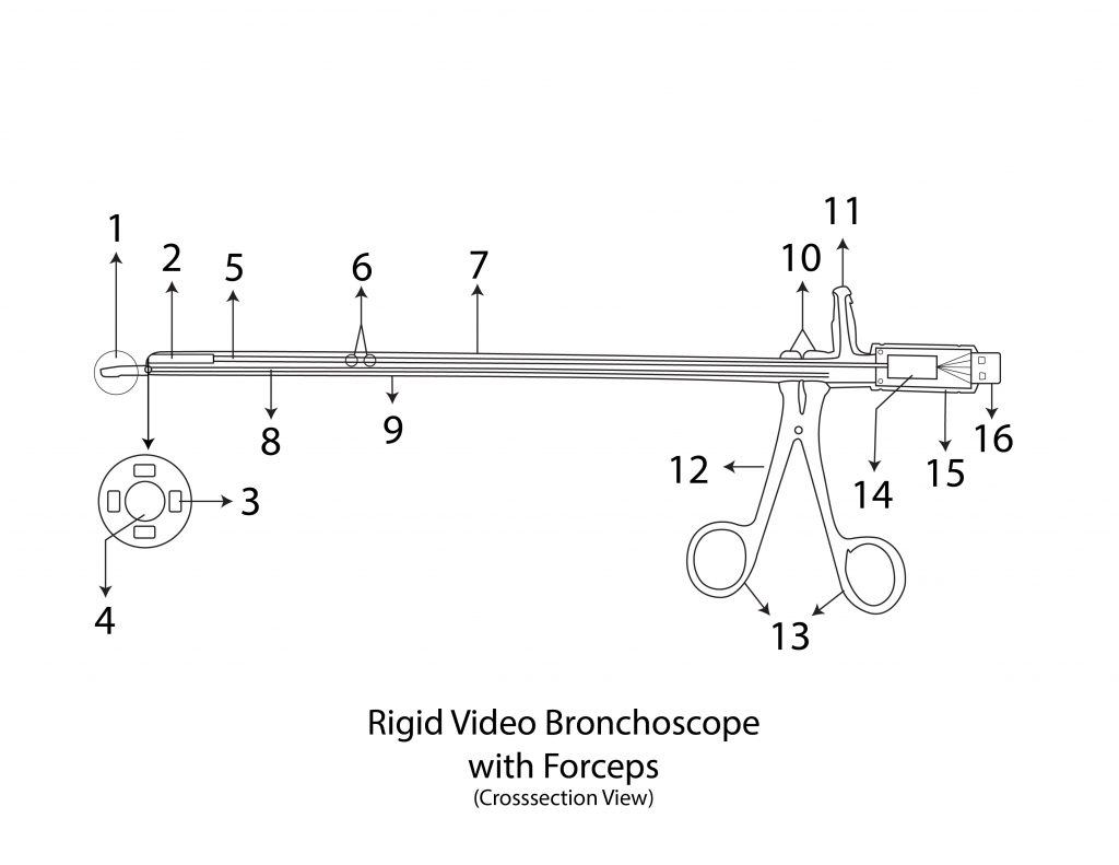
Figure 2 showing the inner cross-sectional view of rigid ventilating video bronchoscope with forceps
1. Peanut holding forceps 2. Video Camera side view 3. LEDs (light emitting diodes) 4. Video Camera front view 5. Electrical wires 6. Holes for ventilation 7. Ventilating channel 8. Wire of forceps 9. Body of forceps 10. Heads of handle 11. Port for ventilation 12. Arm of handle 13. Finger holder of handle 14. Circuit board of camera 15. Holder 16. USB pin
The instrument costs of Rs. 40,000 compared to Rs. 5 Lakhs for the conventional equipment. It makes the instrument the cheapest commercially available instrument in the world. The instrument is unique in design and reported for the first time in the medical literature. The instrument is extremely light weight and weighs only 150 gm making it lightest bronchoscopic assembly compared to 500 gm weight of the conventional instrument with endo camera attached. Heavy instrument puts stress on the surgeon hands and a light assembly maintains finer hand movements for a long time. Bronchoscopic foreign body removal takes only 15 mins with instrument compared to 60 minutes for the conventional. The conventional instruments take a lot of time for its assembly which is saved with this instrument. The ventilating ability makes reduces the risk and operative time dramatically. The instrument can record videos and still pictures very easily for documentation and for medicolegal purpose. The recording by conventional instrument is costly, tedious, and significantly increases weight of assembly due to the endo camera and leads to fatigue of surgeon’s hands and a compromised fine hand movement after some time.
Intended application: The device is used for a variety of diagnostic bronchoscopic procedures such as diagnosis of tracheoesophageal H type fistula, medical lung diseases. It can be used for therapeutic bronchoscopy for removal of bronchial foreign body, biopsy of suspicious lesions, brush biopsy and for bronchial lavage, etc.
Method of using rigid ventilating video bronchoscope with forceps: The instrument is sterilized by Ethylene Oxide gas. Patient is under general anaesthesia. The ventilation port is connected to the anaesthesia machine with tubing. A USB cable connects to the USB pin of the device to display devises such as a mobile phone, Laptop, android tabs or a desktop with a free software installed which is available on Play store. The software now displays and record the video and images. The tip of the instrument is put into the trachea and bronchus of the patient and foreign body is diagnosed. The anesthetist is ventilating the patient through the ventilating port. The foreign body is held by
Class B
Material:316 grade stainless steel
Patent No:201621037874
Packing & storage Condition: The product is stored in a dust free cabinet at 25-40 % centigrade temperature. It is packed with multiple layers of bubble wraps and placed in a metal container of appropriate size and sent by courier
Publication: https://www.ijorl.com/index.php/ijorl/article/view/1452/895
Video quality: https://www.youtube.com/watch?v=x-m9dsdj-Sg
Cost: Rs.40,000